Nobuo Kumano(Former manager of Toshiba nuclear-medicine department) |
2.6 Development of SPECT system
Around 1975, X-ray CT became popular, exerting a deep influence on nuclear medicine. One is an attempt to measure the cross-sectional image with nuclear medicine, and to perform SPECT by using a detector of gammacamera. The other is an attempt of acquire functional information by data processing of nuclear medicine image. This is based on the concept that the morphological diagnosis of human body should be done with high-definition X-ray CT and that nuclear medicine should acquire exclusively functional images. This matter will be mentioned later.
In 1978 and 1979, we conducted basic research where we acquired projection data of a subject who was seated on a rotary chair that was located in front of a gammacamera. Based on this research, we developed a commercial product that rotated a gammacamera detector around a subject to acquire the projection data. Toshiba Corp. (hereinafter referred to as Toshiba) made a joint research with Mie University. We made a gantry that supported two gammacamera detectors of 35 cm in diameter, and announced model GCA-70A in 1980.
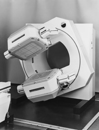 |
| Figure 5. Detector gantry of two square detectors- type SPECT, Toshiba GCA-90A-E2 |
Shimadzu Corp. (hereinafter referred to as Shimadzu) had sold LFOV of Nuclear Chicago (later, Searle, Siemens). They attached two LFOV detectors to their own gantry and sold it as LFOV-E in 1981.
GE started to sell a very simple SPECT system, Maxi Cam. 400T in 1980. In this system, a ring bearing was mounted to the support of counterbalance type gantry. Japanese manufacturers were surprised to see the GE product, because they all had to make a robust gantry to support a heavy detector.
Hitachi Medical Corp. (hereinafter referred to as Hitachi) started to sell RC-1C-1635LF, a multi-purpose gammacamera in 1981. SPECT measurement was possible, and the camera was supported by a counterbalance.
Toshiba sold a SPECT with a square detector mounted, as GCA-90A, in 1982. We mounted two square detectors on the gantry of GCA-70A, and developed a SPECT, GCA-90A-E2 in 1982. Figure 5 shows the gantry of GCA-90A-E2.
2.7 Development of dedicated SPECT
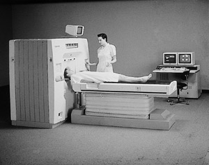 |
| Figure 6. Three detectors-type dedicated SPECT (head mode), Toshiba GCA-9300A |
SPECT measurement was then in wide use for the liver, lung, kidney, heart, whole-body bone. In order to acquire image of higher measurement accuracy, we started development of a dedicated SPECT.
Shimadzu made a joint research with Research Institute for Brain and Blood Vessels Akita, and developed a hybrid ring camera SET 230 capable of SPECT and PET, as well as a head SPECT SET 010, both in 1981.
Hitachi made a joint research with Osaka University, and a head SPECT, SPECT 2000H in 1986.
SPECT must use a collimator, in comparison with PET that was being developed in parallel. For this reason, development of collimator was necessary to increase sensitivity and improve resolution of SPECT measurement.
We delivered GCA-90B-E2 to Kanazawa University. Professor Hisada of that university suggested design of a dedicated SPECT where three detectors were supported in a triangular form like X-ray CT. We called this GCA-9300A and sold it in 1989. Figure 6 shows GCA-9300A.
This dedicated SPECT is a digital gammacamera having three square detectors. The effective field of view 38 cm x 21 cm, slightly smaller than the conventional detector. When the trunk (for example, the heart) of the subject is measured, the sensitivity is 1.5 times the opposing type detector. When the head is measured, a cover is provided so that the subject does not see the detector rotating. For the head application, a fan-beam collimator was especially designed. It has a focus in the rotating direction and parallel holes in the slice direction. It achieved a balanced performance of higher sensitivity and higher special resolution than the conventional parallel hole collimator. Professor Hisada contributed a paper on cerebral blood flow measurement using this device to the annual meeting of SNM (Society of Nuclear Medicine) in 1990. At the highlight lecture on the last day of meeting, Professor Wagner selected the image of cerebral blood flow using HMPAO as the Image of the Year.

2.8 Development of correction technology of detector performance
In 1970s, when a gammacamera was used for planar measurement, the allowable limit was ±10% for the variation of detector sensitivity within the effective field of view. It presented no problem for scintigram reading by gamma ray density distribution. The daily maintenance of detector was performed to keep this level.
At this time, a SPECT was produced in which a detector was rotated around a subject. Toshiba sold GCA-70A in 1981. But, for SPECT measurement, the sensitivity of about ±10% often caused a ring artifact on measurement image. The uniformity data was used and correction was performed using software. The condition of scattered radiation disturbed correction, and caused additional artifact. The non-uniformity of detector was mainly caused by linearity (image distortion) and energy difference at different detection position. The on-line correction, such as Z signal, was necessary for each gamma ray detected. This became possible by the progress of microprocessor, semiconductor memory, incorporated software etc. As a result, the sensitivity improved to ±1.5 to 2.5%. A ring artifact of SPECT was almost removed. With these basic performances corrected, Nuclear Chicago announced ZLC series in 1981.
Toshiba announced GCA-601E SPECT with effective field of view of 35 cm in diameter. It also started to sell a super jumbo digital camera GCA-90A SPECT with a square detector of 50 cm x 35 cm in 1982. It put into a commercial use GCA-90B-E2 SPECT with two square detectors.
Furthermore, we developed technology to prevent deterioration of detector with age. We also developed technology to correct in real time the photo multiplier tube (PMT) sensitivity. This was the main cause of performance change of detectors, when the detector direction changes to the direction of earth magnetism. GE announced Maxi Cam. Autotune ZS in 1983. Toshiba announced SB series of gammacamera in 1989 by incorporating these correction functions and solving the ring artifact problem.
In order to provide the detector and calculation circuit with correction function, we needed a program that is supported by a data processor. For example, the correction data acquired before installation of the system must be converted into the correction data acquired after its installation in the hospital.
Formerly, data was acquired by a gammacamera and sent to a data processor. The emerging trend was a digital gammacamera incorporating the camera and data processor.
2.9 Development of nuclear medicine data processor
Since the age of scintillation scanner, the nuclear medicine image had very smaller number of picture elements than X-ray image. Therefore, the image was processed with a computer even before introduction of a gammacamera. A gammacamera enabled measurement of two-dimensional image. But, the information volume of image did not increase significantly. Instead, they developed measurement technology, such as time data processing, ECG-gated acquisition for cardiac measurement counting etc. This technology developed later into the cardiac measurement for ultrasound diagnosis, X-ray CT and MRI. They also developed sum and subtraction of image data, region of interest processing etc., which were later applied to DR system.
2.9.1 The dawn of data processor development
When we started development of a gammacamera, we already incorporated the concept of a data processing system. However, the initial product was merely two-dimensional multi-channel analyzer. The main specifications were two-dimensional display of image density measured with a gammacamera and its profile display. When Toshiba exhibited a gammacamera GCA-101 at International Congress of Radiology held in 1969, the data processor was also exhibited together.
Subsequently, according as X-ray CT became popular, we feared that if diagnostic imaging by nuclear medicine remains useful only for capture of morphological images, and then its usefulness would decline. Under these circumstances, researchers were expected to develop such a product that aims at functional diagnosis represented by cardiac nuclear medicine.
For that purpose, a data processor of multi-channel analyzer type is not sufficient, because the data processing function is fixed and incapable of change and improvement. An innovative processor should incorporate a general-purpose minicomputer for industry use as a core component, and acquire data from a gammacamera. The data processing software should be developed. Then, the existing processor can be used only by replacement of software. Thus, a new type of nuclear medicine data processor emerged.
In 1970, Toshiba supplied to Kobe University the nuclear medicine image-data-processor DAP-5000 that adopted the minicomputer. In 1973, Toshiba marketed the advanced model DAP-5000N. In 1971, Hitachi marketed the nuclear medicine data processor EDR-4000. In 1972, Shimadzu announced the nuclear medicine data processor Scintipack 200 that used the minicomputer. Shimadzu combined the Scintipack with the gammacamera made by Nuclear Chicago, and developed cardiac analysis software to become a front runner in the market.
2.9.2. Progressive era of nuclear medicine data processor
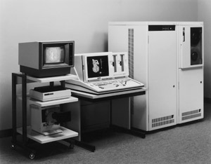 |
| Figure 7 Nuclear medicine data processor Toshiba GMS-55A |
In 1979, Shimadzu marketed the upgraded Scintipack 1200, and established the position of manufacturer of the nuclear medicine data processor. In 1980, Shimadzu developed the Scintipack 70 that had the image memory with an epoch-makingly large capacity as that time, and advertized the high speed of data acquisition and processing.
From 1970, the SPECT using the detector of the gammacamera was commercialized. Data processing as SPECT became necessary to control the detector gantry, to control acquisition of projection data, and to reconstruct SPECT image. In order to perform these SPECT-related processing, the nuclear medicine data processor required the advanced hardware and software.
In 1969, Toshiba started a joint research with Mie University to develop SPECT. Initially, we combined GMS-80A, which was introduced from U.K. under technical license. We found that there was a limit in expandability. In 1981, we developed GMS-55A that used our own minicomputer. Figure 7 shows the appearance of GMS-55A.
This was the nuclear medicine data processor that had large image memory, comparable to Scintipack 70. It used the high resolution display and the function key abundantly.
In the same year of 1981, Shimadzu marketed the Scintipack 2400 with the function in which image processing can be performed in parallel during processing and acquisition of SPECT image.
In the same year of 1981, Hitachi marketed the nuclear medicine data processor HARP. The data processor memorizes the operator's keyboard input procedure. Unskilled operators can handle data processing by using the protocol processing. It was a forerunner product to simplify operation.
The planar measurement by the gammacamera or SPECT measurement acquires only little quantity of the gamma ray. So, the acquisition time ranges from several minutes to several tens of minutes. While acquisition continues, it is possible to process the data that have been acquired. This simultaneous processing function was incorporated at this time into the nuclear medicine data processing. The data processing speed increased, thus shortening the time required for data processing. In 1986, Toshiba marketed GMS-550U that is capable of simultaneous acquisition and processing. It is a stand-alone processor and can be connected to a gammacamera. It was used as a data processor that was incorporated to a digital gammacamera.
2.10 Development of digital gammacamera
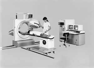 |
| Figure 8 Digital gammacamera Toshiba GCA-601E |
As mentioned above, the correction circuit corrects the performance of detector, and a data processor must support this circuit by means of software. And the function of this data processor was only acquisition, processing, display, recording and storage of conventional gammacamera, and it became insufficient. When a data processor is combined with SPECT, it needs an interface that controls the gantry movement at the time of acquisition. It resulted in the increased information linkage between a camera and a data processor.
Under these circumstances, a data processor computer must control the detector performance and gantry movement and other related job. Individual small tasks are handled by necessary units. As a whole, a computer-controlled gammacamera suits the purpose. The conventional gammacamera measures and sets gamma ray energy (nuclide setup) with switched on the panel. In contrast, the digital camera uses a display, a mouse and a keyboard. Toshiba marketed this type of camera, GCA-601E in 1984, and GCA-901A in 1985. Figure 8 shows GCA-601E.
In 1986, Shimadzu marketed SNR-500R. In 1987, Hitachi marketed RC-150E and RC-135E. The market entered the digital gammacamera age.
In the meantime, Hitachi developed a full digital camera. Digital technology was applied to the output signal from PMT of detector, the calculation circuit for incidence position of gamma ray, and other correction circuits. thus eliminating instability that was likely to occur in analog circuits. As a result, Hitachi marketed RC-1500i in 1991, and RC-2600 in 1992.
3. Development of gammacamera related to my personal history
In 1970 and 1971, Toshiba began to export gammacameras. Personally, I was busy then with arranged introduction for marriage, engagement, Osaka Expo, marriage, etc. In May 1971, Toshiba exported GCA-101 as a No. 1 unit for Europe and I visited Leiden University in the Netherlands for installation. The doctors of university hospital and engineers of our local sales company asked me repeatedly why I did not come together with my newly-wed wife. I found a wide gap in the status level of between Dutch engineers and Japanese (Toshiba) engineers, and I felt the so-called culture shock.
In January 1988, Toshiba achieved production of 1000 units of gammacameras, and held a commemoration ceremony in March. Just several days before that, I went for skiing to Happo-One mountain with Mr. Komatsu and other colleagues. Near the top of mountain, I happened to fall down on the fresh snow. I had a single-lens reflex camera around my neck. The telephoto lens unit hit my body, and broke the left-upper-arm bone condyle. The left shoulder was fixed by gypsum, which covered also the left elbow to the right shoulder. I was unable to wear a suit. I wore a loose cardigan in awkward appearance, and said some greetings. I was in a cold sweat.
If you have engaged in the development, manufacture, sales, servicing etc. of medical devices, then you would feel that your job is worth doing for the society in terms of diagnosis and therapy of diseases. Design and development engineers, like myself, do not use the devices in clinical sites and usually listen to opinion of uses at the site in order to design and develop better products that reflect their opinion. In that sense, engineers must be very sensitive. My seniors always told me that way. It is implicitly expected that our products are used for medical care and the society in general.
We were tied up with a busy routine, such as specifications, cost and delivery. To solve these difficult issues, we must make a great deal of efforts. It was necessary to balance each factor, realize a product, and contribute the benefits of an enterprise. This was an actual situation.
In the spring of 1992, Dr. Yui at Chiba Cancer Center happened to tell me his experience, which made me feel that I was lucky to be an engineer.
He once felt poor health, such as giddiness, and had a cerebral SPECT examination by GCA-9300A. Minute abnormalities were found in a brain blood flow, and he underwent an operation in response to a precision diagnosis. He said, "Without measurement by GCA-9300A such minute abnormalities would have remained undetected, leading more serious condition. The detection would have been delayed, resulting in my prolonged recovery. My life was saved thanks to GCA-9300A."
We had many problems to develop GCA-9300A. The engineers made a great deal of efforts to solve them and succeed in commercial production. I told the above-mentioned episode to all the members concerned and I felt myself that our work was worth doing as a manager of nuclear medicine department.
4. Future of SPECT and PET
In Japan, Mr. Mori at Aloka Co., Ltd. started development of a gammacamera. After that, several companies competed to improve the performance, mainly the special resolution as mentioned here. In 1980s, the digital technology advanced and digital correction was applied to further improve the basic performance of detectors. It resulted in popularization and wide use of SPECT measurement.
However, the market of the gammacamera is screened by the wave of globalization like other medical diagnostic imaging equipment. The companies that develop and sell everything by themselves are decreasing in number. Toshiba combines their own processor with Siemens' camera. Shimadzu combined their own processor with Nuclear Chicago's camera, and now its sells Picker's camera and processor. Initially, Hitachi sold their own processor with Picker's products. Later, it sold a full digital camera of their own development. Most of gammacameras sold in Japan are American products. This is quite regrettable as a former engineer who made a painstaking effort in the early stage of development of gammacamera.
However, in 1970s when development of SPECT started, development of PET started almost at the same time. Both Shimadzu and Hitachi continued that its market is rapidly growing as a clinical PET partly because of abundant supply of FDG, radiopharmaceutical for PET. Besides, PET-CT, a combination with CT, is attracting attention as a system for the purpose of fusion with morphological images and correction of absorption. PET-CT is expected to grow continuously, because it is featured with functional images that can be produced only with nuclear medicine.
Many kinds of radiopharmeceuticals are available for SPECT. Some of them are expected to be further developed exclusively for SPECT. SPECT and PET can and should coexist. We anticipate a steady development of both systems. Formerly, for many years, detectors consisted of a combination of scintillator and PMT. In future, semiconductor detectors will be widely used mainly because of their high energy resolution. But, we still have high barriers to overcome, such as upsizing, cost, stability, etc, and we hope that research and development will continue. We desire that semiconductor detectors will soon be able to contribute to further improvement of performance of both SPECT and PET.
5. Epilogue
In writing this article entitled "Thee development history of gammacamera in Japan," I accessed the website "JIRA Virtual Museum." I referred mainly to pages of "The history of nuclear medicine," but added my comments. I am afraid that some comments are too arbitrary and impartial. Anyway, I have few data and documents available now at hand, and I have relied on my poor memory. I should apologize readers for wrong information and discomforting comments, if any.
In order to further substantiate the content above-mentioned Virtual Museum, readers are kindly requested to provide more information. I have read those pages listing the events chronologically, and found some missing information based on my experience. The missing items cannot be filled with the memory of a single person. If the content is further substantiated by your cooperation, then it will contribute to the future development of medical imaging devices.
The description of PET cyclotron deals with the events only until 2005. The description of nuclear medicine stops at only one item in 2000. The content needs updating.
In this article, I have described mainly gammacamera and its derivative SPECT. I think that PET is also an important item, because it was developed in the same period of SPECT, and development is continuing now. I hope that PET will be described by some other authors in future.
Bibliography
1) History of nuclear medicine: JIRA Virtual Museum, http//www.jira-net.or.jp/vm/chronology_coremedi.html
2) Shigeyasu Kurihara, Nobuo Kumano, Masamichi Katsurada, Eiji Kashio: The gammacamera and its peripheral equipment, Toshiba Review, 26, 2, 1971
3) Nobuo Kumano: Latest nuclear medicine diagnostic equipment, Toshiba Review, 34. 2, pp 108-113 (Showa 54-2) 1979
4) Makoto Kakegawa et al.: Mobile gammacamera GCA-50A, Toshiba Review, 35, 10, pp 812-816, (Showa 55-9) 1980
5) Nobuo Kumano: Nuclear medicine diagnostic equipment, Toshiba Review, 38. 8, pp 691-695, (Showa 58-8),1983
6) Nobuo Kumano: The radioisotope image by the gammacamera, Radioisotopes, 33, 12, 908-915, 1984
7) Nobuo Kumano: Product information [II], Radiology Compendium, Vol. 36, In-vivo nuclear medicine general remarks, 137-143, Nakayama Shoten (1985)
8) Nobuo Kumano: 7. Gammacamera (1) SPECT-- The history from 1981 to 2000--, RADIOISOTOPES, 50, 96S-103S (2001)
9) Nobuharu Yui: My history of nuclear medicine (2001)
10) Nobuo Kumano: Lecture / "The relation of radiation and medicine" 6th session, Radiation and industry, No. 102, 64-71 (2004) |